Contents
Bioimpedance Tomography is a new diagnostic tool that detects bioelectrical signals in a variety of tissues. Its applications in the field of medicine are growing rapidly. This article discusses bioimpedance tomography, image reconstruction algorithms, and physiologic models. It also explores the technology’s clinical applications.
Bioimpedance Tomography
Bioimpedance Tomography (EIT) is a radiation-free method of studying body tissue. It produces images of the human body by measuring changes in impedance across a large area. It also gives detailed information about the cells that make up the human body. Bioimpedance varies depending on the size and orientation of a cell, and its thickness is also a factor.
Clinical applications
Clinical applications of impedance tomography include monitoring of cardiac function and lung perfusion. It has also been used in the management of patients in the critical care setting. It may help monitor regional lung ventilation during surfactant administration and may help minimize the occurrence of volutrauma.
Image reconstruction algorithms
The aim of image reconstruction algorithms for impedance tomography is to reconstruct images from noisy impedance data. This requires solving the inverse problem in a nonlinear fashion and regularizing the results based on a priori knowledge. This thesis presents an approach to solve the problem using neural networks. The approach is based on finite element simulations of the forward problem and is adapted to the inverse problem.
Physiologic models
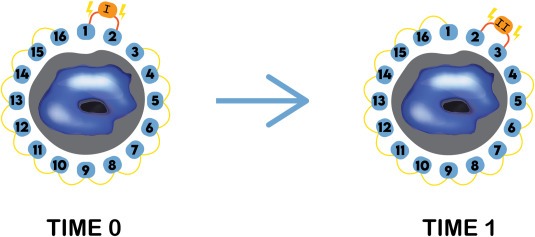
The application of alternating current through two contiguous electrodes is a fundamental method for obtaining Impedance Tomography (EIT) measurements. This method also detects various morphological pathologies of the brain, including cerebral ischemia and hemorrhage. The electrical signal passes through the thorax, following different pathways, depending on the shape of the chest wall and the presence of intrathoracic structures. The impedance changes are then recorded on a computer.
Current-voltage measurements
Current-voltage measurements with impedance-tomography are a powerful method for imaging cellular structures. The technique involves a series of impedance measurements that are transformed into tomographic images using special software. Most systems employ a sensitivity matrix that links the resistivity of each pixel in the image.
Changing thoracic shape
Electrical Impedance Tomography (EIT) is a noninvasive, radiation-free way of studying the thoracic structure. It is particularly useful in monitoring the distribution of ventilation in the lung and preventing ventilator-induced lung injury. However, EIT’s clinical application is limited by the difficulties in interpreting the resulting images. Moreover, because of the inherent inconsistencies between the thoracic shape and the body’s shape, the reconstructed images are prone to errors.
Limitations of the technique
Electrical impedance tomography (EIT) is a non-invasive, radiation-free, and patient-safe diagnostic procedure that provides real-time, continuous monitoring of mechanical properties of the lung. The technology has been around for 30 years, but only recently has it found applications in clinical practice. With improvements in hardware and data processing methods, the field is beginning to see an increased demand for the procedure. However, limitations exist. While most studies have relied on small, heterogeneous groups of patients, more rigorous trials are required to establish its role in everyday clinical practice.


